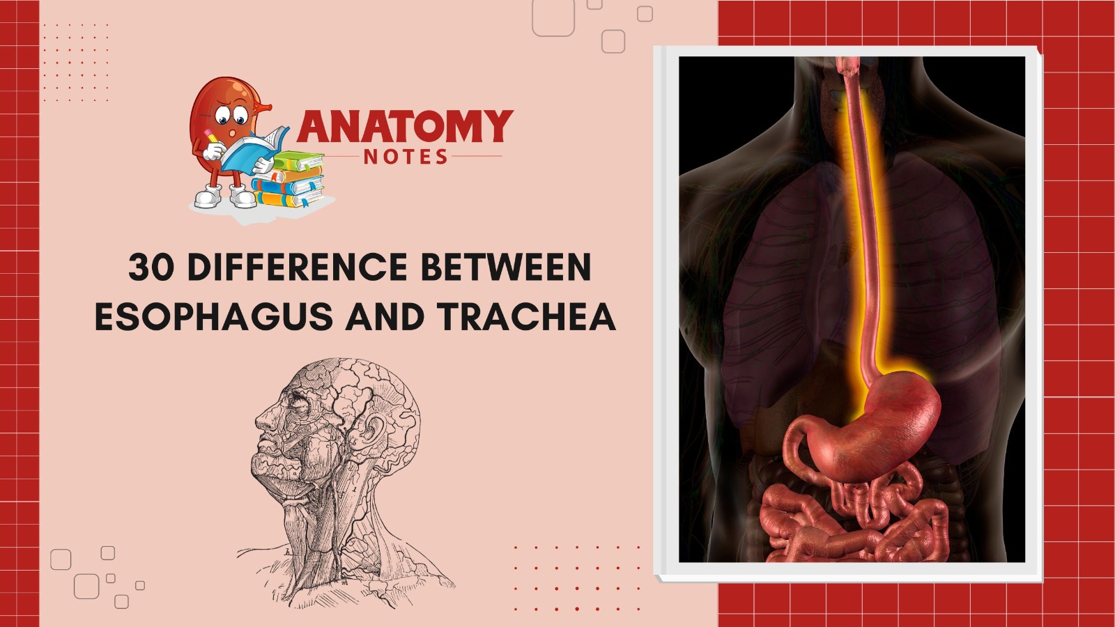
The esophagus and trachea are crucial physiological structures that perform different respiratory and digestive functions. Despite their neck and chest closeness, they differ in various ways. One of the main roles of the esophagus is to transfer ingested food and liquids to the stomach for digestion. Conversely, the trachea is essential to gas exchange by allowing air to travel between the lungs and the outside world.
The smooth muscle and connective tissue esophagus have a multilayer wall that contracts rhythmically to drive ingested particles down. However, cartilage rings support the trachea, preventing collapse while breathing and keeping an open airway. A “windpipe.” These cartilage rings give the trachea its distinctive look.
Linings are another difference. The esophagus’ stratified squamous epithelium protects against food abrasion. Mucus-producing goblet cells and pseudostratified ciliated columnar epithelium line the trachea. Cilia-driven evacuation of foreign particles from this lining protects it.
While the trachea branches into two major bronchi that go to the lungs’ lobes, the esophagus does not branch. This branching distributes air efficiently to lung tissues. The esophagus runs posterior to the trachea, the midline of the body. The anterior trachea makes gas exchange easy. One key distinction is their motions. The esophagus moves food via peristalsis, coordinated muscle contractions. Instead, the trachea coughs and uses cilia to remove mucus and foreign particles.
The esophagus and trachea are close to the neck and chest, yet they have different functions in the digestive and breathing systems. They differ in structure, linings, functions, and movement, demonstrating the body’s sophisticated architecture to control breathing and digestion.
Also Read: Introduction to Cartilage, its formation, structure, and type
Here are 30 differences between the esophagus and trachea:
|
S.No. |
Aspect |
Esophagus |
Trachea |
|
1 |
Location |
In the upper part of the chest, behind the trachea. |
In the front of the neck, below the larynx. |
|
2 |
Function |
Transports food and liquids from the mouth to stomach. |
Conducts air from the larynx to the bronchi and lungs. |
|
3 |
Wall Composition |
Composed of smooth muscle tissue. |
Composed of cartilage rings and smooth muscle tissue. |
|
4 |
Shape |
Tubular, with a muscular wall. |
Tubular, with a rigid cartilaginous structure. |
|
5 |
Lining |
Lined with mucous membrane for food passage. |
Lined with ciliated mucous membrane for air passage. |
|
6 |
Presence of Cilia |
Generally lacks cilia. |
Contains cilia to trap and move mucus and debris. |
|
7 |
Secretions |
Produces mucus for lubrication. |
Produces mucus to trap foreign particles and clean air. |
|
8 |
Food Passage |
Allows food to pass through via peristalsis. |
Does not allow food passage; only air passes through. |
|
9 |
Location in Digestive |
Part of the digestive system. |
Part of the respiratory system. |
|
10 |
Muscular Contractions |
Undergoes rhythmic contractions for swallowing. |
No rhythmic contractions; passive airflow. |
|
11 |
Presence of Glands |
Mucous glands are present for lubrication. |
Mucous glands present for mucus production. |
|
12 |
Sensory Innervation |
Contains sensory nerves for detecting food passage. |
Contains sensory nerves for detecting irritants in air. |
|
13 |
Food Breakdown |
No digestion or breakdown of food occurs. |
No role in food breakdown or digestion. |
|
14 |
Passage of Digestive |
Leads to the stomach. |
Does not lead to the stomach. |
|
15 |
Passage Obstruction |
Can become obstructed by food impactions. |
Obstruction can occur due to foreign objects or swelling. |
|
16 |
Role in Respiration |
No role in respiration. |
Plays a crucial role in the respiratory system. |
|
17 |
Vulnerability to |
Less susceptible to infections and irritations. |
More susceptible to infections and irritations. |
|
18 |
Surgical Procedures |
May require surgery for conditions like GERD. |
May require surgery for conditions like tracheal stenosis. |
|
19 |
Airway Protection |
Does not provide protection for the airway. |
Provides protection for the airway by preventing collapse. |
|
20 |
Diameter |
Larger in diameter than the trachea. |
Smaller in diameter than the esophagus. |
|
21 |
Airway Patency |
No need to maintain airway patency. |
Must maintain airway patency at all times. |
|
22 |
Connection to Stomach |
Connects to the stomach at the lower esophageal sphincter (LES). |
No connection to the stomach. |
|
23 |
Role in Gas Exchange |
Does not participate in gas exchange. |
Participates in gas exchange within the lungs. |
|
24 |
Role in Sound Production |
Has no role in sound production. |
Contributes to voice production by housing vocal cords. |
|
25 |
Vulnerability to Trauma |
Less vulnerable to trauma from external forces. |
More vulnerable to trauma from external forces. |
|
26 |
Role in Speech |
Not involved in speech production. |
Plays a role in speech production. |
|
27 |
Length |
Shorter in length compared to the trachea. |
Longer in length compared to the esophagus. |
|
28 |
Presence of Sphincters |
Contains the lower esophageal sphincter (LES). |
Does not contain sphincters. |
|
29 |
Role in Food Storage |
Does not store food. |
No role in food storage. |
|
30 |
Response to Foreign Bodies |
May lead to choking if foreign objects enter. |
Foreign objects are more likely to be coughed up or expelled. |
Also Read: Muscular System: Muscle types, characteristics & functions
Frequently Asked Questions (FAQS)
Q.1 What are the main functions of the esophagus and trachea?
The esophagus mostly transports food and drinks to the stomach. Peristalsis rhythmic contractions do this. However, the trachea helps air move from the larynx to the lungs. It is essential for respiration, exchanging oxygen and carbon dioxide.
Q.2 Where are the esophagus and trachea located in the body?
The esophagus is behind the trachea and spine. It descends the chest and crosses the diaphragm to reach the stomach. The trachea runs from the larynx to the bronchi in the lungs in front of the esophagus. Its form is maintained by C-shaped cartilage rings.
Q.3 How do esophagus and trachea structures differ?
The esophagus contracts smooth muscle to force food down. The food channel lining is intended to tolerate mechanical stress. In contrast, cartilaginous rings in the trachea maintain the airway open and flexible. Ciliated cells in the trachea transport mucus and debris from the lungs.
Q.4 Do the esophagus and trachea have different protective mechanisms?
Yes, they do. The lower esophageal sphincter, a muscular ring, prevents acid reflux by blocking stomach acid. The flap-like epiglottis covers the trachea during swallowing to keep food and liquids out.
Q,5 What happens if the esophagus or trachea are blocked?
A blockage in the esophagus can cause discomfort, difficulty swallowing, and food stuckness. This can be caused by strictures, tumors, or inflammation. A trachea obstruction can cause coughing, breathing problems, and even death. Airway blockages can be caused by foreign items, edema, or growth.


