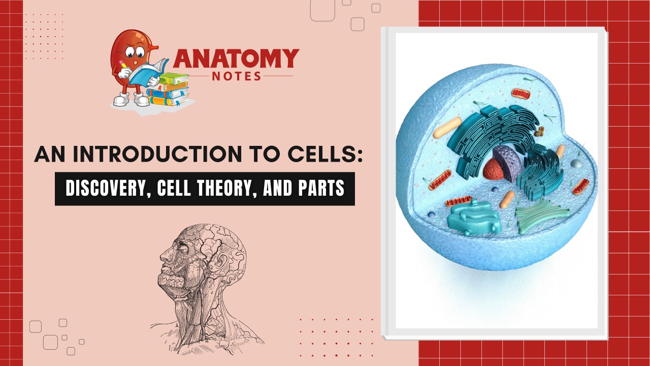
The cells are the fundamental structural and functional units of all living organisms, and anything less than a complete cell has no independent existence.
In 1665, Robert Hooke observed a thin section of cork under a compound microscope, noticed a honey-comb like compartments. He coined the term “Cell”.
In 1674, Anton Von Leeuwenhoek first saw and describe the living cell.
Later, in 1831, Robert Brown discovered the nucleus in the root cells of orchids.
The invention of the microscope & its improvement leading to the electron microscope revealed all the structural details of the cell.
CELL THEORY
In 1838, Matthias Schleiden found that all the plant cells have essentially similar structure and have a cell wall.
In 1839, Theodore Schwann studied different types of animal cells and reported that it had a thin outer layer which is today known as the ‘plasma membrane’.
He also concluded that the presence of the cell wall is a unique character of the plant cells. On the basis of this Schwann proposed the hypothesis that the bodies of animals & plants are composed of cells & its product.
Schleiden and Schwann together formulated this theory. This theory, however, didn’t explain how they were formed.
In 1855, Rudolf Virchow first explained that the cells divided and new cells are formed from pre-existing cells. He modified the hypothesis of Schleiden and Schwann to give the cell theory a final shape.
This theory as understood today is:-
All living organisms are composed of cells and their products.
All cells arise from pre-existing of itself.
Also Read:
Sensory System-Introduction, Organs, and Functions
https://anatomynotes.org/sensory-organs/sensory-system-introduction-organs-and-functions/
CELL COMPONENTS
Plasma membrane:
It is the outermost, it is a thin layer, transparent, elastic & semi-permeable membrane. It helps in the exchange of materials between the cytoplasm and extracellular fluid.
Nucleus:
The nucleus also called the director of the cell, is the most important part of it which directs and controls all the cellular functions
Cell Organelles:
The eukaryotic cell consists of following cell organelles:
- Endoplasmic Reticulum
- Ribosomes
- Golgi apparatus
- Mitochondria
- Lysosomes
- Fibrils
- Microtubules
- Centrioles
- Inclusions
ENDOPLASMIC RETICULUM – Endoplasmic Reticulum (ER) is a network of tiny tubular structures scattered in the cytoplasm. The ER bearing Ribosomes on their surface is called Rough Endoplasmic Reticulum (RER). In the absence of Ribosomes, they appear smooth and are called Smooth Endoplasmic Reticulum (SER).RER is actively involved in protein synthesis and secretion. The SER is the major site for lipid synthesis & lipid-like steroidal hormones.
RIBOSOMES – These are granular structures, composed of RNA and proteins & they are surrounded by any membrane. The eukaryotic Ribosomes are the 80S while prokaryotic Ribosomes are 70S. These are the sites of protein synthesis.
GOLGI BODIES – They consist of many flat, disc-shaped sacs or cisternae of 0.5 1 micron diameters, stacked parallel to each other. It involved cell secretion, the formation of hormones, biosynthesis of glycolipids & glycoproteins.
MITOCHONDRIA – It is a double membrane-bound structure with the outer membrane and inner membrane dividing its lumen into two compartments. They are called powerhouses of the cell as these are sites of ATP formation.
LYSOSOMES – These are vesicular structures of the cytoplasm that are involved in intra-cellular digestive activities; contain all types of hydrolytic enzymes, also known as the suicidal bag of the cell.
FIBRILS – They are present in the various cells. It helps to maintain the cell shape and is also responsible for the contractility as they are present in the muscle fibers.
MICROTUBLES – They become part of mitotic spindles in dividing cells.
CENTRIOLES – These are the pair of short rod-shaped bodies found adjacent to the nucleus lying at right angles to each other. centrioles also give rise to cilia or flagella.
CILIA & FLAGELLA – Cilia & Flagella are microscopic, hair or thread-like motile structures present extra-cellularly but originate intra-cellularly and help in locomotion, feeding, circulation, etc.
INCLUSIONS – These are pigments like melanin or lipofuscin, storage granules such as glycogen and fat, and secretion granules.
Learn more:
Frequently Asked Questions (FAQs)
What are human cells and their functions?
Human cells are the basic building blocks of the human body. They carry out a variety of functions including energy production, nutrient absorption, waste removal, and the maintenance and repair of tissues and organs. They also play a crucial role in the immune response, communication between different parts of the body, and the transmission of genetic information.
What type are human cells?
There are many different types of human cells, each with their own unique structure and function. Some of the most common types of human cells include muscle cells, nerve cells, skin cells, blood cells, and immune cells. Within each of these categories, there are further subtypes of cells that specialize in different functions. For example, blood cells can be further divided into red blood cells, white blood cells, and platelets.
What color is a nucleus?
The nucleus of a human cell is not colored. It is a small, usually spherical organelle that is surrounded by a double membrane and contains genetic material in the form of DNA. While the nucleus itself is not colored, it can be stained with dyes such as hematoxylin and eosin to highlight its structure and location within a cell. The color of the stain can vary depending on the specific dye used and the staining protocol, but typically ranges from shades of purple to pink.
What color is DNA?
DNA (deoxyribonucleic acid), the genetic material that is contained within the nucleus of a human cell, does not have a specific color. DNA is a complex molecule that does not absorb or emit visible light, and therefore it does not have any inherent color. However, scientists often use dyes to visualize DNA in the lab, and these dyes can give the DNA a color. For example, ethidium bromide, a common DNA stain, fluoresces orange-red when it binds to DNA, which can make the DNA appear orange or red under ultraviolet light.
What is the smallest human cell?
The smallest human cell is the sperm cell, also known as spermatozoon. Sperm cells are produced in the male reproductive organs, specifically the testes, and are essential for sexual reproduction. They are typically around 50 micrometers in length and have a unique structure that enables them to swim towards the egg and fertilize it. While sperm cells are the smallest human cells, they are still highly specialized and contain many different structures and organelles.
What are 90% of skin cells?
The vast majority of skin cells are keratinocytes, which make up about 90% of all cells in the epidermis, the outermost layer of the skin. Keratinocytes are responsible for producing and maintaining the skin’s protective barrier, which helps to prevent the entry of harmful substances and the loss of moisture. These cells also produce a tough, fibrous protein called keratin, which gives the skin its strength and elasticity. In addition to keratinocytes, the skin also contains other types of cells, such as melanocytes, which produce the pigment melanin, and immune cells that help to protect against infection.


