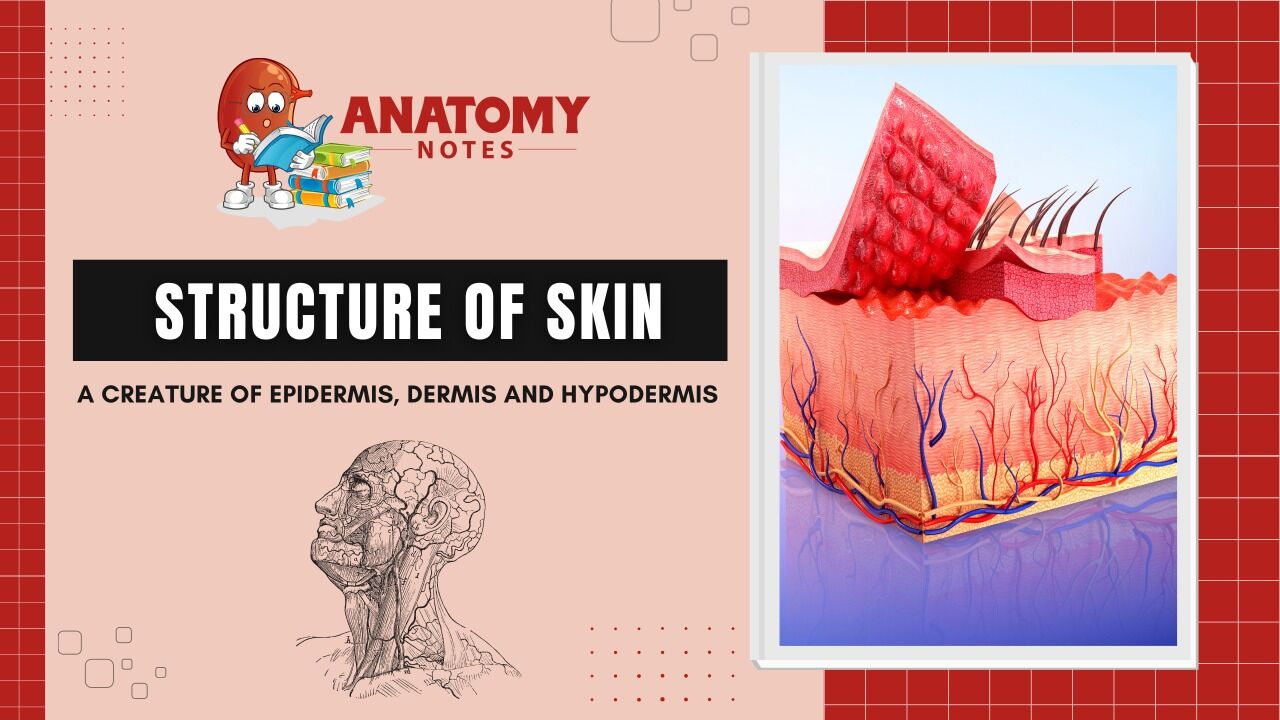
The skin is made up of three major layers:
- Epidermis – It is made up of closely packed epithelial cells.
- Dermis – It is made up of dense, irregular connective tissue that includes blood vessels, hair follicles, sweat glands, and other structures.
- Hypodermis – It is composed mainly of loose connective and fatty tissues.
Epidermis – The Topper of the Skin
The epidermis is the topmost layer of skin that can touch, see and feel. It is made up of closely packed epithelial tissue.
The epidermis has no blood supply and it is nourished by diffused oxygen from surrounding air.
The thickness of the epidermis is approximately 0.1mm. It acts as a protective layer as it protects the entering of pathogens.
The epithelial layer is further divided into five layers which are mentioned below:-
Stratum corneum
- The stratum corneum is the most superficial layer of the epidermis and is the layer exposed to the outside environment.
- The increased keratinization of the cells in this layer gives it its name. There are usually 15 to 30 layers of cells in the stratum corneum.
- This dry, dead layer helps prevent the penetration of microbes and the dehydration of underlying tissues and provides mechanical protection against abrasion for the more delicate, underlying layers.
- Cells in this layer are shed periodically.
Stratum lucidum
- The stratum lucidum is a smooth, seemingly translucent layer of the epidermis located just above the stratum granulosum and below the stratum corneum.
- This thin layer of cells is found only in the thick skin of the palms, soles, and digits.
- The keratinocytes that compose the stratum lucidum are dead and flattened.
- These cells are densely packed with eleiden, a clear protein rich in lipids, derived from keratohyalin, which gives these cells their transparent (i.e., lucid) appearance and provides a barrier to water.
Stratum granulosum
- The stratum granulosum has a grainy appearance due to further changes to the keratinocytes as they are pushed from the stratum spinosum.
- The cells (three to five layers deep) become flatter, their cell membranes thicken, and they generate large amounts of the proteins keratin, which is fibrous, and keratohyalin, which accumulates as lamellar granules within the cells.
- These two proteins make up the bulk of the keratinocyte mass in the stratum granulosum and give the layer its grainy appearance.
- The nuclei and other cell organelles disintegrate as the cells die, leaving behind the keratin, keratohyalin, and cell membranes that will form the stratum lucidum, the stratum corneum, and the accessory structures of hair and nails.
Stratum spinosum
- The stratum spinosum is spiny in appearance due to the protruding cell processes that join the cells via a structure called a desmosome.
- The desmosomes interlock with each other and strengthen the bond between the cells.
- It is interesting to note that the “spiny” nature of this layer is an artifact of the staining process.
- Unstained epidermis samples do not exhibit this characteristic appearance.
- The stratum spinosum is composed of eight to 10 layers of keratinocytes, formed as a result of cell division in the stratum basale
Stratum germinativum or stratum basale
- It is the deepest epidermal layer and attaches the epidermis to the basal lamina, below which lie the layers of the dermis.
Dermis – The First Runner Up
The dermis is the core of the integumentary system. It contains blood and lymph vessels, nerves, and other structures, such as hair follicles and sweat glands.
The dermis is made of two layers of connective tissue that compose an interconnected mesh of elastin and collagenous fibers, produced by fibroblasts.
It has two layers which are listed below –
Papillary layer
- The papillary layer is made of loose, areolar connective tissue, which means the collagen and elastin fibers of this layer form a loose mesh.
- This superficial layer of the dermis projects into the stratum basale of the epidermis to form finger-like dermal papillae.
- Within the papillary layer are fibroblasts, a small number of fat cells (adipocytes), and an abundance of small blood vessels.
- In addition, the papillary layer contains phagocytes, defensive cells that help fight bacteria or other infections that have breached the skin.
- This layer also contains lymphatic capillaries, nerve fibers, and touch receptors called the Meissner corpuscles
Reticular layer
- It is composed of dense, irregular connective tissue. It is well vascularized and has a rich sensory and sympathetic nerve supply.
- The reticular layer appears reticulated (net-like) due to a tight meshwork of fibers.
- Elastin fibers provide some elasticity to the skin, enabling movement.
- Collagen fibers provide structure and tensile strength, with strands of collagen extending into both the papillary layer and the hypodermis.
- In addition, collagen binds water to keep the skin hydrated.
- Collagen injections and Retin-A creams help restore skin turgor by either introducing collagen externally or stimulating blood flow and repair of the dermis, respectively.
Hypodermis – Finally The Inner One
The hypodermis is also known as the subcutaneous layer or superficial fascia.
It is a layer that directly lies below the dermis and serves to connect the skin to the underlying fascia (fibrous tissue) of the bones and muscles.
It is not strictly a part of the skin, although the border between the hypodermis and dermis can be difficult to distinguish.
The hypodermis consists of well-vascularized, loose, areolar connective tissue and adipose tissue, which functions as a mode of fat storage and provides insulation and cushioning for the integument.
Frequently Asked Questions (FAQs)
What is skin and its types?
The skin is the body’s largest organ and serves as a safeguarding barrier that separates the internal organs from the external environment. There are three main types of skin: oily, dry, and normal. Skin can also be categorized based on its sensitivity and susceptibility to conditions such as acne, rosacea, and eczema.
What are the 7 structures of the skin?
The 7 structures of the skin are:
- Epidermis – the outermost layer of the skin
- Dermis – the layer beneath the epidermis that contains nerves, blood vessels, and hair follicles
- Hypodermis or subcutaneous tissue – the deepest layer of the skin that contains fat and connective tissue
- Hair follicles – tiny cavities within the dermis that produce hair
- Sebaceous glands – small glands within the dermis that produce sebum, an oily substance that helps lubricate the skin and hair
- Sweat glands – small glands within the dermis that produce sweat
- Nerves and blood vessels – found throughout the skin to provide nourishment and sensation.
How many cells are in the skin?
The number of cells in the skin varies depending on the size and thickness of the skin, as well as other factors. However, on average, there are approximately 19 million skin cells in every square inch of the human body. This means that the total number of skin cells in an average-sized adult is estimated to be around 10 trillion.
What is skin structure function?
The skin performs various tasks, which include:
- Protection: The skin acts as a barrier to protect the body from harmful external factors, such as UV radiation, bacteria, and chemicals.
- Sensation: The skin contains nerve endings that allow us to sense touch, pressure, pain, and temperature.
- Thermoregulation: The skin helps to regulate body temperature by producing sweat when the body is too hot and constricting blood vessels when the body is too cold.
- Vitamin D synthesis: The skin produces vitamin D when exposed to sunlight, which is important for bone health.
- Excretion: The skin excretes sweat and other waste products through sweat glands, helping to eliminate toxins from the body.
What is the functional layer of skin?
The functional layer of skin is the dermis, which is the layer beneath the outermost layer of skin, known as the epidermis. The dermis is responsible for providing the skin with strength, flexibility, and elasticity. It contains collagen and elastin fibers, as well as blood vessels, hair follicles, and sweat and sebaceous glands. The dermis also contains nerve endings that allow us to sense touch, pressure, pain, and temperature. In addition, it plays an important role in regulating body temperature, as well as in wound healing and scar formation.
What are the 3 layers of skin and their structures?
The skin is composed of three main layers, each with its own unique structure and function. The three layers of the skin, listed in order from the outermost layer to the innermost layer, are:
- Epidermis: This is the outermost layer of the skin, composed mainly of keratinocytes, which are cells that produce the protein keratin. The epidermis also contains melanocytes, which produce the pigment melanin that gives skin its color, and Langerhans cells, which are part of the immune system.
- Dermis: This is the layer beneath the epidermis, composed mainly of collagen and elastin fibers, as well as blood vessels, hair follicles, and sweat and sebaceous glands. The dermis provides the skin with strength, flexibility, and elasticity, and contains nerve endings that allow us to sense touch, pressure, pain, and temperature.
- Subcutaneous tissue (or hypodermis): This is the deepest layer of the skin, composed mainly of adipose tissue and connective tissue. The subcutaneous tissue helps to insulate and cushion the body, as well as provide a reserve of energy.