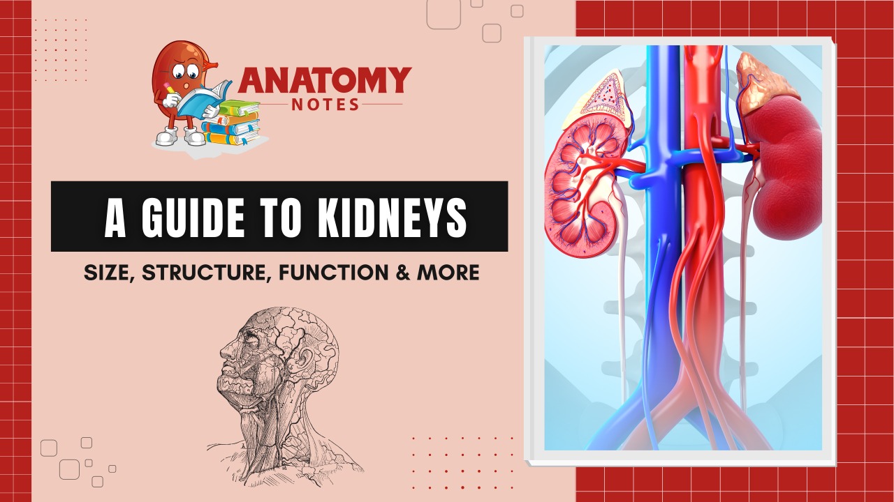
Kidneys are present in pair inside the human body, which are located on the left and right side of the abdomen. kidneys help in to excrete the end products of metabolism and the excess water.
Location– The kidneys located in the posterior abdomen. They lie retroperitoneally i.e. behind the peritoneum, either side of the vertebral column.
Also Read: Microscopic Structure Of Skeleton Muscles
https://anatomynotes.org/muscular-system/microscopic-structure-of-skeleton-muscles/
If we see Superiorly, then we will find that they lie in the upper border of the 12th thoracic vertebrae and if we see Inferiorly, then they are level with the third lumbar vertebrae. There is a little bit difference between the left and right kidney that is left kidney is slightly higher than the right kidney due to the presence of massive liver in the right hypochondrium.
Size and Shape- They are basically bean-shaped organs. In adults, each kidney generally has 11cm length, 6cm breadth, and 3cm height. The height of the left kidney is 1.5cm more than the right one. Each of them weighs about 150g in men and 135g in the female. They also occur in lobules form in the fetus and newborn, generally it has 12 lobules. But in adults, these lobules fused to present a smooth surface.
Structure of kidney
The kidney is bean-shaped with two side i.e. outer convex side called as reddish-brown cortex and inner concave side called renal hilus where the renal vein, artery, and ureter are present.
The renal capsule is a thin connective tissue which surrounds each kidney and maintains the shape of it and helps to protect the inner tissues of it.
The renal cortex is the outer layer which is present inside the renal capsule. Renal cortex is soft, dense and a vascular tissue. Under the renal capsule a layer is present which is known as renal medulla, which consist many renal pyramids. These pyramids are in the cone shaped with apices pointing toward the center of kidney.
Each and every apex of the renal pyramid is connected to a minor calyx and minor calyx is a hollow collecting tube for urine which merge and form three major calyces that also merge into the renal pelvis at the Hilus. And from where, urine drains out into the ureter.
The following are the parts of it which perform different-different types functions:
Also Read: The Urinary System-Introduction, Functions and Anatomy
https://anatomynotes.org/urinary-system/the-urinary-system-introductionfunctions-and-anatomy/
- Renal Hilus – It is the depression which is present near the center of the concave side of kidney where renal vein and ureter is absent and the renal artery enters into the kidney.
- Renal capsule -it is a smooth and transparent membrane which surrounds the kidney and help to protect and maintain the shape of kidney. it is also surrounded by fatty tissue which prevent any damage to kidney.
- Renal cortex- it is the outer reddish part of the kidney with smooth texture with bowman’s capsule, glomeruli, proximal and distal convoluted tubules and blood vessels.
- Renal medulla- It is the inner striated red brown part of the kidney.
- Renal pyramids – it is striped, triangular lien under the medulla which are made up of straight tubules and corresponding blood vessels.
- Renal pelvis – A funnel shaped cavity that receives urine which drained from the collecting ducts and papillary ducts.
- Renal artery – these blood vessels delivers oxygenated blood to the kidney. Renal artery enters the kidney through the hilus and divides into smaller arteries, which further separates into afferent arterioles that serve each of the nephron.
- Renal vein – These are the blood vessels which delivers the de-oxygenated blood to the kidney and return it to the systemic circulation.
- Interlobular artery – these blood vessels helps in to deliver oxygenated blood to glomerular capillaries in high temperature.
- Interlobular vein – these blood vessels helps in to recieves de-oxygenated blood to which is drain from glomeruli and the Henle’s loop
- Kidney nephron – it is the functional unit if kidney which performs main function of kidney. it is present in millions in each kidney.
- Collecting duct – it plays role in collecting urine and drains into papillary ducts, minor calyx and major calyx and in last into the ureter and urinary bladder.
- Ureter – it is the tube-like structure which helps to pass urine from the kidney to the urinary bladder.
Functions of Kidneys
The urinary system depends on proper excretory organ structure and performance. a number of these core actions include:
- Excretes waste: They eliminates toxins, urea, and excess salts And some organic compound which could be a nitrogen-based waste matter of cell metabolism that’s made within the liver and transported by the blood to the kidneys.
- Maintains water balance: They facilitate maintain water and balance within the body. They react to changes within the water level, which can increase or decrease throughout the day.
- Regulates pressure of blood: They facilitate regulate blood pressure by the production of angiotensin, a substance that constricts the blood vessels and signals the body to retain water and sodium when pressure is low.
- Regulates red blood cells: They produce a hormone i.e. erythropoietin, which stimulates our bone marrow to supply additional red blood cells once when the body doesn’t get enough gas.
- Regulates acid levels: Acids are the product of metabolism. The kidneys facilitate maintain the correct acid-base equilibrium to stay the body healthy.
Organs Associated with Kidneys
There are different groups of structures those associated with kidneys
Organs associated with Right kidney
- The adrenal gland which lies superiorly.
- The right lobe of the liver, the duodenum, and the hepatic flexure of the colon, they all are lies anteriorly.
- The diaphragm, and the muscles of the posterior abdominal wall.
Organs associated with Left kidney
- The left adrenal lies superiorly
- The spleen, pancreas, jejunum, and splenic abdominal wall, they all are lies anteriorly.
- The diaphragm, and the muscles of the posterior abdominal wall.
Learn more: ALL MEDICAL STUFF
Frequently Asked Questions (FAQs)
How to improve kidney function?
Some ways to improve kidney function include drinking plenty of water, reducing salt intake, maintaining a healthy diet, exercising regularly, avoiding smoking and excessive alcohol consumption, managing blood pressure and blood sugar levels, and taking prescribed medications as directed by a healthcare professional.
Can the consumption of a large amount of water have a positive effect on the health of your kidneys?
Drinking an adequate amount of water is essential for maintaining healthy kidneys. Water helps to flush out toxins and waste products from the kidneys, preventing the formation of kidney stones and other kidney problems. However, drinking excessive amounts of water can be harmful and put a strain on the kidneys, so it’s important to stay hydrated in moderation. A general guideline is to drink at least 8 glasses (64 ounces) of water per day, but individual needs may vary depending on factors like age, activity level, and overall health.
What causes kidney damage?
There are numerous factors that could lead to kidney impairment, such as:
- Diabetes: High blood sugar levels can damage the blood vessels in the kidneys, impairing their ability to filter waste products from the blood.
- High blood pressure: Elevated blood pressure can damage the blood vessels in the kidneys, reducing their ability to function properly.
- Autoimmune diseases: Conditions like lupus and vasculitis can cause inflammation in the kidneys, leading to damage over time.
- Infections: Certain infections, such as pyelonephritis, can cause kidney damage if left untreated.
- Medications: Some medications, such as nonsteroidal anti-inflammatory drugs (NSAIDs) and certain antibiotics, can cause kidney damage if taken in high doses or over a long period of time.
- Genetic factors: Some forms of kidney disease are hereditary and can be passed down through families.
- Trauma: Injury to the kidneys from accidents or sports injuries can cause kidney damage.
- Aging: As we age, our kidneys may become less efficient at filtering waste products from the blood, leading to kidney damage over time.
What are the main structures of the kidney?
The main structures of the kidney include the renal cortex, renal medulla, renal pelvis, renal artery and vein, nephrons, collecting ducts, and ureters. The renal cortex and medulla contain millions of nephrons, the functional units of the kidneys, which filter waste products from the blood and produce urine. The urine is then transported to the renal pelvis through the collecting ducts and exits the kidney through the ureters.
Where is the kidney structure?
The kidneys are a pair of bean-shaped organs located in the back of the abdomen, on either side of the spine. They are situated just below the rib cage, with the right kidney slightly lower than the left to make room for the liver. The kidneys receive blood from the renal artery, filter it through the nephrons, and return the cleaned blood to circulation through the renal vein. The urine produced by the kidneys is transported to the bladder through tubes called ureters.
What is the unit of kidney?
The nephron is the term used to refer to the basic structural and functional unit of the kidney. Each kidney contains millions of nephrons, which are responsible for filtering waste products and excess fluid from the blood and producing urine. The nephron consists of several structures, including the glomerulus, Bowman’s capsule, proximal and distal tubules, and collecting ducts. The glomerulus filters the blood, and the filtrate then passes through the tubules, where various substances are reabsorbed or secreted depending on the body’s needs. The resulting urine is then transported to the bladder for excretion.


